Key Takeaways
- 3D models provide an interactive way to study the digestive system, enhancing understanding and retention.
- Detailed exploration of the digestive system begins with the mouth and ends at the rectum, with each organ playing a critical role.
- 3DTotal’s 3D models offer a step-by-step guide to accessing and utilizing features such as rotation, zoom, and clickable layers for an in-depth learning experience.
- Visual learning with 3D models caters to different learning styles and can be especially beneficial for young learners and medical professionals.
- Incorporating 3D models into educational curricula and patient education promotes engagement and simplifies complex concepts.
Starting the Anatomical Journey
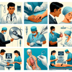
Embarking on an anatomical journey can be as thrilling as it is enlightening. Whether you’re a seasoned medical professional or a curious student, the intricate workings of the human body never cease to amaze. And what better way to delve into the marvels of human anatomy than with 3D models? These tools aren’t just visually engaging; they’re a bridge to deeper understanding.
The Role of 3D Models in Learning Anatomy
Imagine being able to explore the nooks and crannies of the digestive system without a single incision. That’s the power of 3D models. They break down barriers, offering a hands-on experience that textbooks alone can’t provide. By turning, flipping, and dissecting these models digitally, learners can visualize spatial relationships and complex structures, leading to ‘aha’ moments that stick.
But it’s not just about the visuals. It’s about interaction. 3D models respond to your curiosity, allowing you to peel back layers and reveal the hidden secrets of the body’s inner workings. This dynamic form of learning caters to a variety of styles, accommodating those who learn best by doing.
Highlights of 3DTotal’s Digestive System Models
3DTotal’s digestive system models stand out for their meticulous detail and user-friendly design. They’re crafted with the user in mind, ensuring that whether you’re guiding a patient through their digestive health journey or mastering the material for an exam, you have a tool that’s both accurate and accessible.
These models don’t just show you the digestive system; they guide you through it. With each click, you’re able to uncover another layer, another function, another piece of the puzzle. And with each discovery, your understanding deepens. It’s an interactive experience that transforms learning from passive to active.
Navigating 3DTotal’s Guided 3D Models
Diving into 3DTotal’s guided 3D models is akin to stepping into a virtual lab where the human digestive system is laid out in front of you, ready for exploration. With intuitive controls and detailed imagery, these models are a gateway to a deeper understanding of human anatomy.
Step-by-Step Guide: Accessing the Models
First things first, to access these comprehensive 3D models, you’ll need to navigate to 3DTotal’s website. Here’s how you can get started:
- Visit the 3DTotal’s official website and head to the educational section.
- Select the ‘Digestive System’ from the list of anatomical models.
- Register or log in to your account to gain full access to the models.
- Once logged in, you can select the digestive system model to begin your exploration.
It’s that simple. Now, with the model at your fingertips, you’re ready to dive into the intricacies of the digestive tract.
Interactive Model Features: Rotate, Zoom, and Explore
3DTotal’s models are designed with interactivity at their core. Here are some features that make these models an invaluable learning tool:
- Rotate: Get a 360-degree view by rotating the model. This allows you to view the digestive system from every angle, ensuring a comprehensive understanding of its layout.
- Zoom: Zoom in and out to focus on specific organs or structures. Want a closer look at the villi of the small intestine? Zoom in to see every detail.
- Explore: Navigate through the different parts of the digestive system by clicking on the organ of interest. This feature allows for a focused study on each part.
With these features, you can control your learning experience and focus on the areas that interest you the most or those you find most challenging.
Understanding Anatomical Structures with Clickable Layers
One of the standout features of 3DTotal’s models is the ability to interact with different layers of the digestive system. By clicking through these layers, you can see how each part connects and works together.
For example, you might start with the outermost layer to see the overall shape and position of the digestive organs. Then, you can peel back this layer to reveal the muscular structure beneath, and then go even deeper to explore the blood vessels and nerves that supply these organs.
This layered approach not only helps in memorizing the anatomy but also provides insights into the functional aspects of the digestive system. It’s a powerful way to learn how the body’s systems are interconnected.
Leveraging 3D Models for Effective Learning
3D models are more than just a technological novelty; they’re a cornerstone of modern anatomy education. By leveraging these tools, students and medical professionals can gain a dynamic and lasting understanding of complex anatomical structures.
Visual Learning: Benefits of 3D Anatomical Representation
Visual learning is essential in the field of anatomy, and 3D models are a testament to this. The benefits are clear:
- They cater to visual learners who need to see concepts in action to understand them fully.
- They provide a spatial understanding that 2D images cannot, which is crucial for grasping the layout of the digestive system.
- They allow for repeated exposure and interaction, which is key to retention and recall.
Therefore, by engaging with 3D models, learners can visualize and remember anatomical details more effectively than through traditional study methods alone.
Reinforcing Knowledge: How to Use Models for Study and Review
To get the most out of 3D models, it’s important to integrate them into your study routine in a way that reinforces your knowledge. Here are some tips to help you do just that:
- Start by exploring the entire model to get a sense of the digestive system’s layout.
- Focus on one organ at a time, using the zoom and rotate functions to examine it from all angles.
- Use the clickable layers to understand how each organ functions within the system.
- Regularly revisit the models to reinforce your memory, as repetition is key to learning.
By following these steps, you’ll find that complex anatomical concepts become clearer and more manageable.
For Educators: Incorporating 3D Models into Your Curriculum
If you’re an educator, integrating 3D models into your curriculum can revolutionize the way you teach anatomy. Here’s how you can bring these tools into your classroom:
- Use 3D models as a visual aid during lectures to illustrate complex concepts.
- Assign interactive model exploration as homework to reinforce classroom learning.
- Encourage students to use models during group study sessions for collaborative learning.
- Incorporate model quizzes and assessments to test students’ knowledge in a practical context.
These strategies can enhance engagement, understanding, and enthusiasm for the subject matter.
Engaging with the Wonders of the Digestive System
Beyond the practicalities of learning and teaching, 3D models invite us to engage with the wonders of the human body in a way that fosters curiosity and respect for its complexities.
Seeing Beyond the Textbook: Models as a Window into the Human Body
3D models offer a window into the human body that textbooks simply can’t match. They bring the static images of a page to life, allowing learners to witness the dynamic nature of the digestive system in action.
This experiential form of learning not only educates but inspires, sparking a sense of wonder that drives the pursuit of knowledge and the desire to understand the intricacies of human health.
Making Complex Concepts Accessible: Simplified Model Explanations for All Ages
The beauty of 3D models lies in their ability to distill complex anatomical concepts into digestible, interactive experiences. With 3DTotal’s models, every detail of the digestive system is simplified, making it accessible to learners of all ages. By breaking down the system into clear, manageable sections, these models serve as an invaluable resource for demystifying the inner workings of our bodies.
The Fascination of Internal Processes: Inspiring Future Biologists and Doctors
There’s a certain magic in seeing the internal processes of the human body come to life. For young minds pondering a future in biology or medicine, 3D models can light the spark of inspiration. They provide a glimpse into a future career, offering a tangible connection to the abstract concepts encountered in textbooks. It’s this kind of engagement that can inspire the next generation of healthcare professionals.
FAQs
In the journey through anatomy with 3D models, questions are bound to arise. Let’s address some of the most common inquiries to ensure that everyone can make the most of these incredible learning tools.
How detailed are 3DTotal’s digestive system models?
3DTotal’s models are meticulously detailed, offering a comprehensive view of the digestive system. From the salivary glands in the mouth to the villi lining the small intestine, every aspect is rendered with precision, providing a depth of understanding that rivals hands-on dissection.
Can I access 3DTotal’s models on my mobile device?
Yes, accessibility is key, and 3DTotal’s models are designed to be used across various devices, including mobile phones and tablets. This means you can take your learning on the go, whether you’re commuting, between classes, or in a clinical setting.
What are the main parts of the digestive system that can be explored with 3D models?
The digestive system is a complex network, and 3D models cover it in its entirety. You can explore the mouth, salivary glands, esophagus, stomach, liver, pancreas, gallbladder, small intestine, large intestine, and rectum. Each model offers detailed views of the organs, tissues, and even the cellular structures that make up these components.
How can 3D models transform the understanding of the digestive process?
3D models transform learning by offering an immersive experience. They allow you to witness the journey of food as it becomes fuel for our bodies. By interacting with each part of the system, you can see how the organs work together, how nutrients are absorbed, and how waste is expelled. This holistic view fosters a deeper appreciation and understanding of human physiology.
Are 3DTotal’s 3D models suitable for all age groups?
Absolutely. 3DTotal’s models are designed to be user-friendly and are suitable for anyone with an interest in learning about the human body. The models are particularly beneficial for young students who can manipulate and explore the digestive system in a way that’s both fun and educational.
Furthermore, the models can be adapted to suit various levels of complexity, making them just as useful for medical students and professionals who require a more detailed exploration of the digestive system.
Can 3D models be used as tools for patient education in healthcare settings?
Yes, they can, and they are incredibly effective. 3D models provide a visual aid that can help patients understand their conditions, treatments, and the importance of digestive health. By seeing a clear representation of what’s happening inside their bodies, patients can feel more empowered and involved in their healthcare decisions.
This visual approach can also alleviate anxiety and improve patient compliance with treatment plans, as they gain a better understanding of the rationale behind medical advice.
What tips do you have for integrating 3D anatomical models into study routines?
To effectively integrate 3D models into your study routine, consider the following tips:
- Set specific goals for each study session, such as focusing on a particular section of the digestive system.
- Incorporate the models early in your learning process to build a strong foundational understanding.
- Use the models in conjunction with other study materials, such as flashcards or quizzes, to reinforce learning.
- Take advantage of the interactive features, like dissection and layering, to deepen your comprehension of each organ’s function and structure.
- Regularly review the models to maintain your knowledge and help commit information to long-term memory.
By following these strategies, you’ll find that 3D anatomical models become an indispensable part of your learning toolkit, paving the way for a more engaging and effective study experience.