Key Takeaways:
- 3D cardiac surgery models offer unprecedented precision in pre-surgical planning.
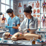
- These models enhance surgical training, providing hands-on experience without risk.
- Customized patient-specific models lead to improved surgical outcomes and patient safety.
- Technological advancements are pushing the boundaries of what’s possible in cardiac care.
- Despite their benefits, 3D cardiac models face financial and technical challenges.
The Cutting Edge of Heart Care: 3D Cardiac Surgery Models
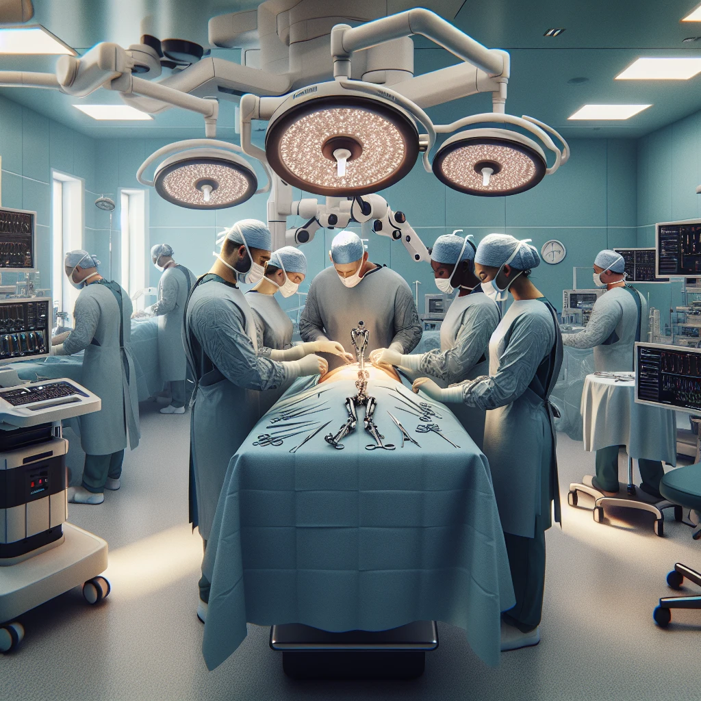
Imagine stepping into an operating room where the heart you’re about to operate on is no stranger. You’ve seen every vein, every valve, and every possible complication, not on a screen, but in your hands. This is the reality that 3D cardiac surgery models bring to the table—literally. These models are game-changers, transforming the way surgeons prepare for and perform heart surgeries.
Revolutionizing Cardiac Surgery with 3D Modeling Technology
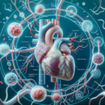
When it comes to heart surgery, precision is not just a goal—it’s a necessity. 3D modeling technology is the surgeon’s ally, offering a detailed roadmap of the patient’s heart. It’s like having a GPS during surgery; you know exactly where to go and what to avoid.
But how do we create these models? It starts with high-resolution imaging techniques such as CT scans or MRIs. These images are then used to create a digital 3D model of the patient’s heart, which can be printed using specialized materials that mimic human tissue. This process not only helps visualize the heart’s structure but also allows surgeons to plan and practice the procedure, reducing the likelihood of surprises during the actual surgery.
Take the case of a young boy with a complex congenital heart defect. Traditional imaging gave a partial picture, but it was the 3D printed model that revealed a previously unseen blood vessel that could have led to complications during surgery. This level of insight is invaluable.
Precision Planning: The Role of 3D Models in Pre-Surgical Procedures
Pre-surgical planning with 3D models is like doing a trial run of a play before opening night. Surgeons can rehearse complex procedures, anticipate challenges, and refine their strategies. This meticulous preparation leads to shorter operation times, less anesthesia, and overall, a safer surgical experience for the patient.
3D Printing in Congenital Heart Surgery
Children with congenital heart defects are the most poignant beneficiaries of 3D printing technology. Their tiny, often uniquely malformed hearts make surgeries especially challenging. 3D models allow surgeons to understand and navigate these complexities with a level of detail that was previously unthinkable.
For instance, in surgeries to correct defects like Tetralogy of Fallot, which involves multiple structural issues within the heart, 3D models provide a tangible, multi-dimensional view. Surgeons can hold the heart in their hands, plan the repair of each defect, and ensure that the delicate tissues are treated with the utmost care.
In one heartwarming story, a little girl’s life was saved thanks to a 3D model. The surgical team discovered an extra muscle bundle inside her heart that wasn’t visible in traditional scans. Without the 3D model, the surgery could have had a very different outcome.
Advancing Treatment with Patient-Specific Models
Every heart is unique, and 3D models celebrate this individuality. They are tailored to the patient’s specific anatomy, providing a personalized approach that enhances the precision of each cut and stitch. This custom fit not only improves the surgery itself but also aids in the patient’s recovery, as the interventions are as minimally invasive as possible.
For surgeons, these models are a bit like dress rehearsals. They can simulate different approaches, find the best route for their instruments, and even predict the heart’s behavior during surgery. This level of preparation is unprecedented and is quickly becoming the gold standard in cardiac care.
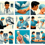
3D Print Technology: From Imaging to Implementation
The journey from imaging to implementation is a marvel of modern medicine. The process begins with the acquisition of detailed images of the heart, which are then converted into a digital 3D model. This model is meticulously reviewed and refined by a team of specialists, ensuring that it accurately represents the patient’s anatomy.
Once the digital model is perfected, it’s time for the 3D printer to work its magic. Using materials that mimic human tissue, the printer creates a physical model of the heart. This model isn’t just a static replica; it’s a dynamic tool that can be manipulated, cut, and sutured just like real tissue.
Hands-On Surgical Training Enhanced by 3D Models
Training for heart surgery has traditionally relied on textbooks, videos, and if you’re lucky, observation. But with 3D models, trainees can roll up their sleeves and dive into the anatomy hands-on. It’s an interactive learning experience that accelerates the development of surgical skills and confidence.
These models are particularly beneficial for practicing procedures that are rare or complex. Trainees can repeatedly perform the surgery on the model, honing their technique in a no-risk environment. This repetition is crucial for muscle memory and decision-making skills, which are vital in the high-stakes world of cardiac surgery.
Educational Benefits of 3D Printed Cardiac Models
3D printed cardiac models are not just tools for experienced surgeons; they’re also powerful educational resources. They bridge the gap between theory and practice, providing a tactile learning experience that is far more engaging than traditional methods.
- Models facilitate a deeper understanding of cardiac anatomy and pathology.
- They allow for interactive learning, where students can dissect and explore without consequences.
- Models enable the visualization of complex spatial relationships within the heart.
- They encourage collaborative learning, as students and teachers can discuss and manipulate the heart together.
- These models can be used to simulate a wide range of scenarios, from routine procedures to rare and complex surgeries.
By incorporating 3D models into medical education, we’re preparing the next generation of surgeons with a level of hands-on experience that was once only possible through years of observation and practice.
Real-World Skills: Impact of 3D Models on Surgical Proficiency
The impact of 3D models on surgical proficiency is profound. They allow surgeons to transition from theory to practice with a confidence that can only come from experience. It’s one thing to read about a heart valve replacement; it’s another to have practiced the procedure on a model that mimics the real thing.
These models also offer a safe space for seasoned surgeons to master new techniques. The field of cardiac surgery is constantly evolving, and 3D models provide a platform for continuous learning and skill enhancement.
Surgeons who train with 3D models are better equipped to handle the unexpected. They’ve ‘seen’ more hearts, more variations, and more potential complications before ever making an incision on a living patient. The result? Faster response times, more effective problem-solving, and ultimately, better patient outcomes.
Let’s not forget the psychological benefits. There’s a confidence that comes from preparation, and 3D models provide that in spades. Surgeons who have practiced with these models approach the operating table with a level of calm and assurance that only comes from thorough preparation.
Case Studies: 3D Models in Action
It’s one thing to talk about the potential of 3D cardiac models, but let’s look at how they’ve actually impacted surgeries. Real-world cases where 3D models have been the difference between success and uncertainty.
Victory Over Ventricular Defects
Consider a case where a young patient presented with a ventricular septal defect (VSD), a hole in the wall separating the heart’s two lower chambers. Traditionally, this condition can be challenging to repair due to the complexity of the surrounding structures and the risk of damaging the heart’s electrical system.
Using a 3D model, the surgical team was able to plan and practice the repair. They knew exactly where to place sutures to close the defect without affecting the surrounding tissue. The surgery was a success, and the patient’s recovery was swift, thanks to the precision afforded by the 3D model.
Conquering Complex Congenital Anomalies
In another instance, a baby was born with a rare congenital heart anomaly that left surgeons puzzled. The traditional imaging was inconclusive, and the risk of going into surgery blind was high.
With a 3D printed model, the surgical team could explore the anomaly from all angles. They discovered routes for repair that would have been impossible to visualize with 2D images alone. The model allowed for a successful surgery that was less invasive and had a significantly reduced risk of complications.
Stories like these underscore the transformative power of 3D cardiac models. They’re not just tools; they’re lifelines, turning what was once uncertain into something tangible and manageable.
In both cases, the 3D models didn’t just assist in the surgical process; they changed the trajectory of these patients’ lives. They represent a leap forward in cardiac care, where the heart’s secrets are revealed before surgery begins, allowing for repairs that are precise, personalized, and profoundly effective.
As we continue to harness the power of 3D printing and modeling in cardiac surgery, we’re not just improving outcomes. We’re reshaping the very nature of surgical education and patient care, one heart at a time.
Procedural Innovations: Specificities of 3D-Guided Surgeries
Now, let’s delve into the specifics of how 3D models are guiding some of the most intricate cardiac procedures. The level of detail provided by these models allows for a degree of innovation and precision that was previously unattainable, particularly in surgeries like mitral valve repair and arterial switch operations.
3D Techniques in Mitral Valve Repair
The mitral valve, with its complex anatomy, has always posed a challenge for repair. But with 3D models, surgeons can now plan and execute repairs with a clarity that ensures both the functionality and longevity of the valve. The model allows the surgeon to assess the valve’s defects, plan the repair, and even test the durability of the repair before the patient is on the table.
For example, a surgeon was able to use a 3D model to simulate the placement of artificial chords in a patient with mitral valve prolapse. This preoperative rehearsal allowed for a precise and successful repair, reducing the patient’s time under anesthesia and improving recovery time.
Arterial Switch Operation Precision with 3D Assistance
Arterial switch operations are complex procedures performed on infants born with transposition of the great arteries. The success of this surgery hinges on the precise reconnection of the arteries to the correct chambers of the heart. With 3D models, surgeons can visualize and practice this delicate procedure, ensuring that the arteries are reconnected without tension or twisting.
Such precision was instrumental in the case of a newborn, where the 3D model allowed the surgical team to identify the best approach for detaching and reattaching the arteries, significantly reducing the risk of postoperative complications.
Optimizing Surgical Outcomes with 3D Cardiac Models
3D cardiac models are not just about improving the surgical experience; they’re about optimizing outcomes for patients. By providing a detailed preview of the surgical landscape, these models help surgeons minimize intraoperative risks and enhance patient safety and recovery trajectories.
Assessment and Reduction of Intraoperative Risks
One of the most significant advantages of 3D models is their ability to help surgeons identify and mitigate potential risks before the surgery even begins. By practicing on the model, surgeons can foresee and avoid complications, leading to a smoother operation and less time on the operating table.
- Models reveal anatomical anomalies that may not be evident in 2D images.
- They allow for the testing of different surgical approaches to find the safest one.
- Surgeons can anticipate and prepare for potential bleeding sites.
- Models provide a clear understanding of the spatial relationship between structures, reducing the risk of accidental damage.
- They enable the surgical team to plan for equipment and support needs specific to the case.
By reducing intraoperative risks, 3D models contribute to a decrease in the likelihood of postoperative complications and the need for additional surgeries.
Enhancing Patient Safety and Recovery Trajectories
When it comes to recovery, every minute of surgery and every incision matters. 3D models help streamline the surgical process, leading to less invasive procedures and, consequently, faster recovery times. Patients benefit from reduced pain, lower risk of infection, and quicker return to normal activities.
Moreover, the precision afforded by 3D models often results in better functional outcomes, such as improved heart function and less need for long-term medications. This is not only beneficial for the patient’s health but also contributes to a reduction in healthcare costs associated with prolonged hospital stays and additional treatments.
Consider a patient who underwent a complex aortic valve replacement. Thanks to the 3D model, the surgical team could minimize the size of the incision and accurately place the new valve, leading to a remarkable recovery. The patient was able to leave the hospital sooner and with less postoperative pain than typically expected.
As we continue to integrate 3D models into cardiac surgery, we are setting new standards for patient care. These models are more than just tools; they are the embodiment of a commitment to excellence in surgical practice and patient outcomes. With each surgery, we are not only repairing hearts but also improving lives, one beat at a time.
The Reality of 3D Models: Limitations and Progression
While the benefits of 3D cardiac models are undeniable, it’s important to recognize the challenges that come with this innovative technology. Like any pioneering field, there are hurdles to clear before these models can become commonplace in every cardiac operating room.
Financial and Technical Barriers
The creation of 3D cardiac models is not without its costs. The technology requires significant investment in high-quality printers, materials, and training. Moreover, the process of converting medical images into printable models demands a level of technical expertise that is not always readily available. These barriers can make the widespread adoption of 3D models a challenge, particularly in resource-limited settings.
- High costs of 3D printers and materials can be prohibitive for some institutions.
- Technical expertise is required to create accurate and functional models.
- There is a need for specialized software and hardware, which may not be accessible to all.
- Time investment for model preparation and printing can be significant.
- Insurance reimbursement for the use of 3D models is not yet standardized.
Despite these challenges, the push for more accessible and affordable 3D printing solutions continues. Innovators and healthcare professionals alike are working to develop cost-effective materials and streamline the production process to bring this technology within reach of more patients and surgeons.
The Future: Overcoming Current Challenges in 3D Modeling
Looking ahead, the future of 3D cardiac models is bright. As the technology advances, we can expect to see reductions in costs and improvements in accessibility. Efforts are underway to create open-source software and collaborative platforms that will make it easier for medical professionals to produce and share 3D models. Additionally, ongoing research into new materials and printing techniques promises to further enhance the quality and realism of these models.
- Development of open-source software to democratize access to 3D modeling.
- Research into new, more cost-effective materials for printing.
- Improvements in printing speed and resolution.
- Increased collaboration between medical institutions to share knowledge and resources.
- Standardization of insurance coverage for 3D modeling in surgical planning.
As these developments unfold, the potential of 3D cardiac models will continue to expand, paving the way for even more innovative uses in cardiac care.
Emerging Trends and Future Developments in 3D Cardiac Surgery
The field of 3D cardiac surgery is not standing still. Emerging trends and future developments are set to push the boundaries of what’s possible, offering new hope and possibilities for patients with heart conditions.
Additive Manufacturing: The Next Frontier in Cardiac Care
Additive manufacturing, more commonly known as 3D printing, is evolving rapidly. The next frontier in cardiac care involves bio-printing, where living cells are used as ‘ink’ to create tissue or even whole organs. This advancement could revolutionize transplant medicine and open up new avenues for treating heart disease.
Imagine a future where a patient’s own cells are used to print a new heart valve that is a perfect match for their body, eliminating the risk of rejection and the need for lifelong medication.
While this future is not yet a reality, research in bio-printing is progressing, and it’s only a matter of time before such procedures are part of standard care.
Integration of Virtual Reality and AI in 3D Cardiac Procedures
The integration of virtual reality (VR) and artificial intelligence (AI) into 3D cardiac procedures is another exciting trend. VR can provide an immersive environment for planning and practicing surgeries, while AI can analyze vast amounts of data to predict outcomes and recommend the best surgical approaches.
- Virtual reality simulations for immersive surgical planning and training.
- Artificial intelligence algorithms to optimize surgical strategies and predict patient outcomes.
- Combining 3D models with augmented reality (AR) for real-time guidance during surgery.
- Machine learning to improve the accuracy and efficiency of 3D model creation.
- AI-driven analytics to personalize patient care and improve long-term health management.
These technologies are not just futuristic concepts; they are being developed and tested right now. As they mature, they will undoubtedly become integral components of cardiac surgery, enhancing the capabilities of surgeons and the care received by patients.
The journey of 3D cardiac models from a novel idea to a surgical staple is a testament to the power of innovation in healthcare. By overcoming current limitations and embracing emerging trends, we are on the cusp of a new era in cardiac surgery—one that promises greater precision, personalization, and improved outcomes for patients around the world. As we continue to push the envelope, the heart of the matter remains clear: technology, when thoughtfully applied, has the power to save lives and change the narrative of heart disease.
Frequently Asked Questions (FAQ)
How Do 3D Cardiac Surgery Models Improve Preoperative Planning?
3D cardiac surgery models are a bit like having a crystal ball. They give surgeons a glimpse into the future of the operation, revealing the heart’s unique anatomy in a tangible way. This allows for meticulous planning, where every cut and suture can be strategized in advance. The models enable the surgical team to identify potential obstacles and devise a clear plan of action, which can reduce operating time and enhance patient safety.
Can All Cardiac Surgeries Benefit from 3D Modeling?
While 3D modeling has a broad range of applications, not every cardiac surgery will require such advanced planning. However, for complex cases—particularly those involving congenital defects, intricate valve repairs, or abnormal anatomy—3D models can be incredibly beneficial. They are also invaluable for educational purposes, helping new surgeons understand the nuances of cardiac anatomy and surgical techniques.
What Is the Accuracy of 3D Printed Heart Models?
The accuracy of 3D printed heart models is quite remarkable. They are created from high-resolution imaging data, which means they can replicate the patient’s heart with great precision. The level of detail in these models allows surgeons to see structures that might not be as clear in 2D images, providing a more complete understanding of the heart’s anatomy. However, the precision also depends on the quality of the imaging and printing technology used.
How Are 3D Models Used in Surgical Training?
3D models are revolutionizing surgical training by providing a hands-on experience that was previously impossible. Trainees can practice procedures on these models, which helps them develop a tactile understanding of the heart’s anatomy and the skills needed for surgery. This practical experience is crucial for building confidence and competence in the operating room.
In conclusion, 3D cardiac surgery models are transforming the landscape of heart surgery. They offer a new level of precision in preoperative planning, enhance surgical training, and have the potential to improve patient outcomes. As the technology continues to evolve, it’s likely that we’ll see even more innovative applications that will further advance the field of cardiac care.